Abdominal x ray tutorial
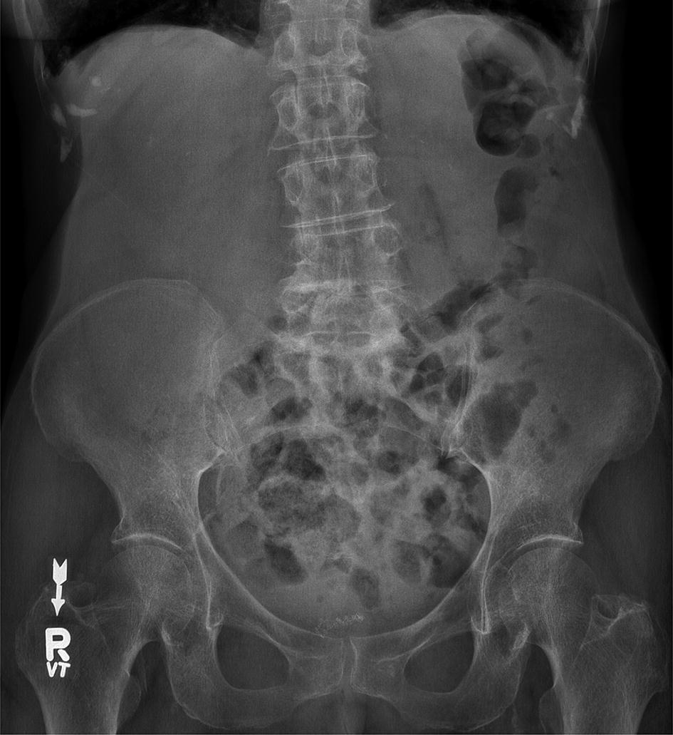
Abdominal X Ray Tutorial. Full assessment includes a check of patient data image quality and checking for artifact and abnormal calcification. It integrated radiological images including X-ray computed tomography and magnetic resonance imaging plus clinical correlations and self-evaluation. The slice thickness is 25 mm. Learn a structured approach to interpreting X-rays.
 Tutorial 10 The Abdominal Radiograph Springerlink From link.springer.com
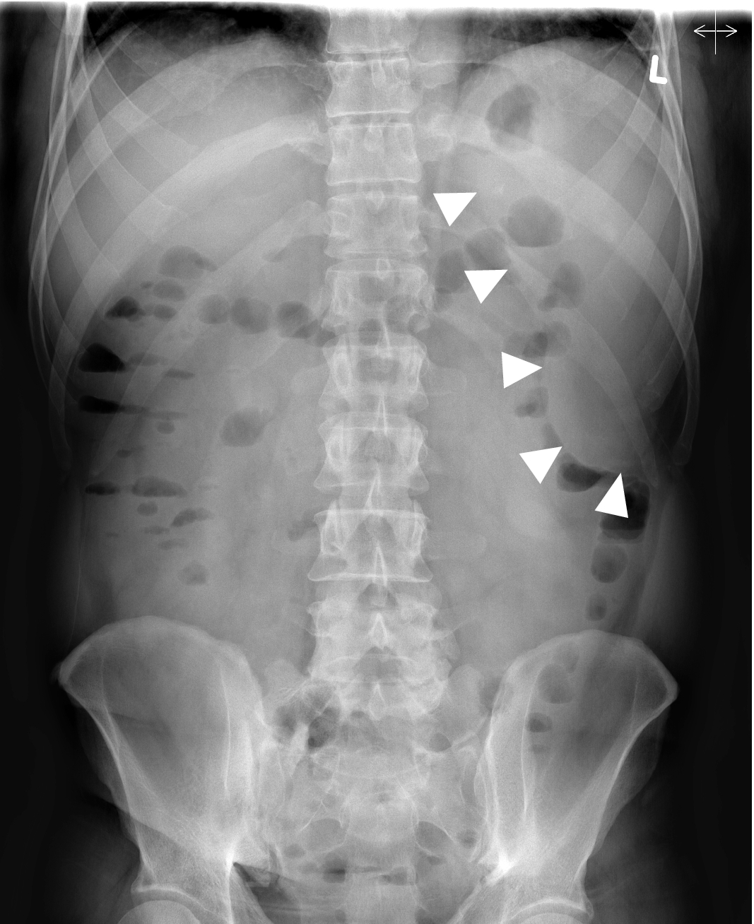
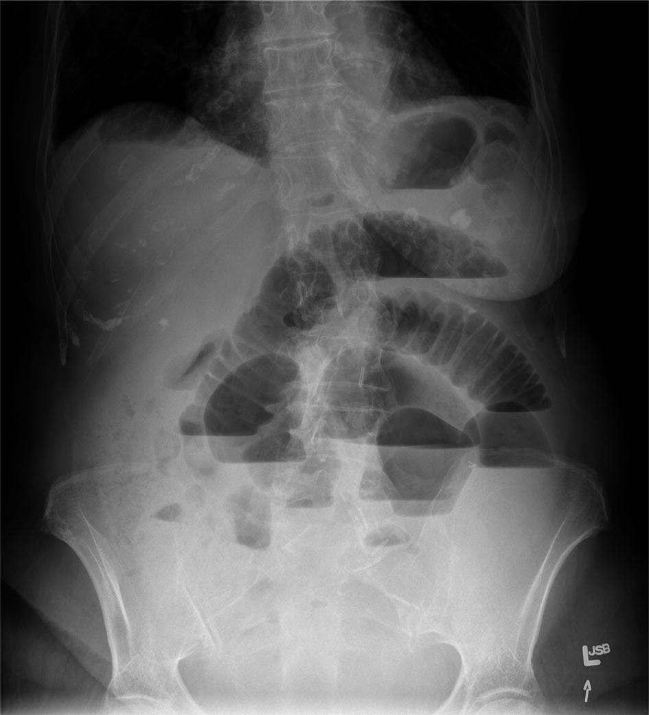
But as you can see from the images above you cannot reliably look for air under the diaphragm in an AXR and thus a CXR is. Typical abdominal X-ray features of small bowel obstruction include dilation of the small bowel 3cm diameter and much more prominent valvulae conniventes creating a coiled-spring appearance. Adhesions are the most common cause of small bowel obstruction in the developed world accounting for 75 of all cases. It integrated radiological images including X-ray computed tomography and magnetic resonance imaging plus clinical correlations and self-evaluation. Whether x-ray is supine or erect for fluid and gas levels correct orientation RightLeft Location of bowel small central large peripheral. A US doctor answered Learn more.
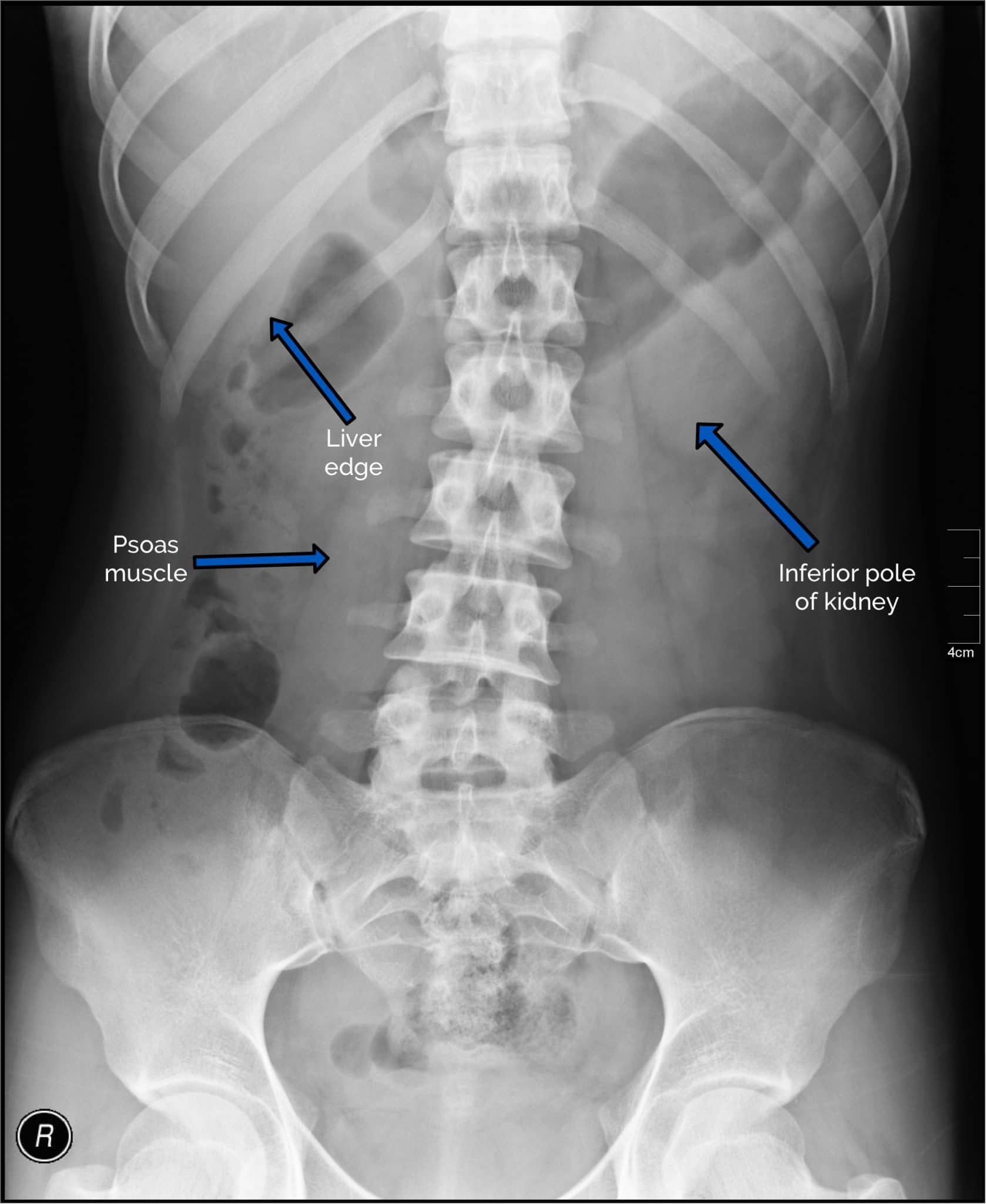
Abdominal X-ray Soft tissue organs and structures of the abdomen Bony structures of the abdomen.
How many rad are in a pelvic and abdominal x-ray. Abdominal X-Rays Tutorial download report. Obesity Obesity interfered with seven out of every 1000 abdominal ultrasounds in 1989. X-rays use beams of energy that pass through body tissues onto a special film and make a picture. It integrated radiological images including X-ray computed tomography and magnetic resonance imaging plus clinical correlations and self-evaluation. Whether x-ray is supine or erect for fluid and gas levels correct orientation RightLeft Location of bowel small central large peripheral.
 Source: slideplayer.com
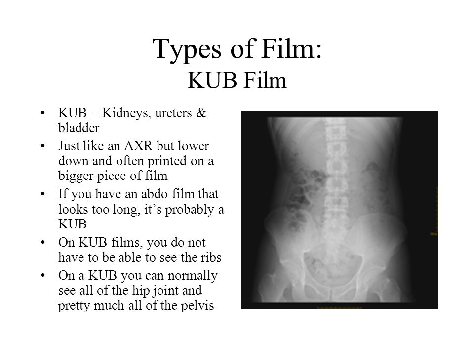
Approach to AXR Bowel gas pattern Extraluminal air Soft tissue masses Calcifications 4. This tutorial will discuss these steps. X-rays of the belly may be done to check the area for causes of abdominal pain. Typical abdominal X-ray features of small bowel obstruction include dilation of the small bowel 3cm diameter and much more prominent valvulae conniventes creating a coiled-spring appearance. Abdominal X-Ray - Small bowel obstruction - Small bowel obstruction can be identified by the dilated loops of centrally placed bowel with the venae commitantes circular bands of muscle that span the entire width of the bowel.
 Source: undergradimaging.pressbooks.com
X-rays of the belly may be done to check the area for causes of abdominal pain. Bone and metal show up as white on X-rays. Adhesions are the most common cause of small bowel obstruction in the developed world accounting for 75 of all cases. Abdominal X-Rays Tutorial EKhalili Pouya 2018. Radiology and medical imaging tutorials for medical students and allied health care professionals.
 Source: pinterest.com
Abdominal X-Rays Tutorial download report. This involves assessment of the bowel gas pattern soft tissue structures and bones. Tutorials also cover acute CT brain. This is a CT of the Abdomen and Pelvis Enterography protocol. X-rays of the belly may be done to check the area for causes of abdominal pain.
 Source: link.springer.com
Learn about abdomen x-ray anatomy. This tutorial will discuss these steps. Because of the difference in X-ray absorption by air and soft tissues the intestinal structures intestinal air can be differentiated from their surroundings. Tutorials covering chest X-ray abdominal X-ray and trauma X-ray interpretation. Abdominal X-Rays Tutorial download report.
 Source: youtube.com
REM is the term. Full assessment includes a check of patient data image quality and checking for artifact and abnormal calcification. Used for occupational or exposure rad for absorbed dose. As a consequence of stomach or bowel perforation. It is performed with a higher radiation dose and larger dose of IV contrast which helps to evaluate subtle areas of bowel inflammation.
 Source: slideplayer.com
In the ramge of 1 to 10 rem. Abdominal X ray 1. In the ramge of 1 to 10 rem. It can also be done to find an object that has been swallowed or to look for a. This is a higher quality study than a standard CT.
 Source: geekymedics.com
Tutorial on sytematic assessment of the abdominal x-ray. It is performed with a higher radiation dose and larger dose of IV contrast which helps to evaluate subtle areas of bowel inflammation. Abdominal X-Rays Tutorial download report. Abdominal X ray 1. REM is the term.
 Source: youtube.com
The tutorial was a self-administered PowerPoint that guided students through principles of abdominal anatomy. As a consequence of stomach or bowel perforation. X-rays use beams of energy that pass through body tissues onto a special film and make a picture. Doctors in 147 specialties are here to answer your questions or offer you advice. The stomach is in the left upper quadrant and is visible when it is filled with air.
 Source: slideplayer.com
X-rays of the belly may be done to check the area for causes of abdominal pain. X-rays use beams of energy that pass through body tissues onto a special film and make a picture. Lightbulb moment a moment of sudden inspiration revelation or recognition 3. Help by adding tags. Kidney and urinary bladder stones and gallstones.
 Source: youtube.com
Because of the difference in X-ray absorption by air and soft tissues the intestinal structures intestinal air can be differentiated from their surroundings. A system for reporting an abdominal X-Ray. X-rays of the belly may be done to check the area for causes of abdominal pain. 2 Air in the abdomen Air rises to the top when there is pneumoperitoneum eg. Abdominal X-Rays Tutorial EKhalili Pouya 2018.
 Source: youtube.com
Abdominal X-Rays Tutorial download report. A system for reporting an abdominal X-Ray. Tutorial on sytematic assessment of the abdominal x-ray. As a consequence of stomach or bowel perforation. Help by adding tags.
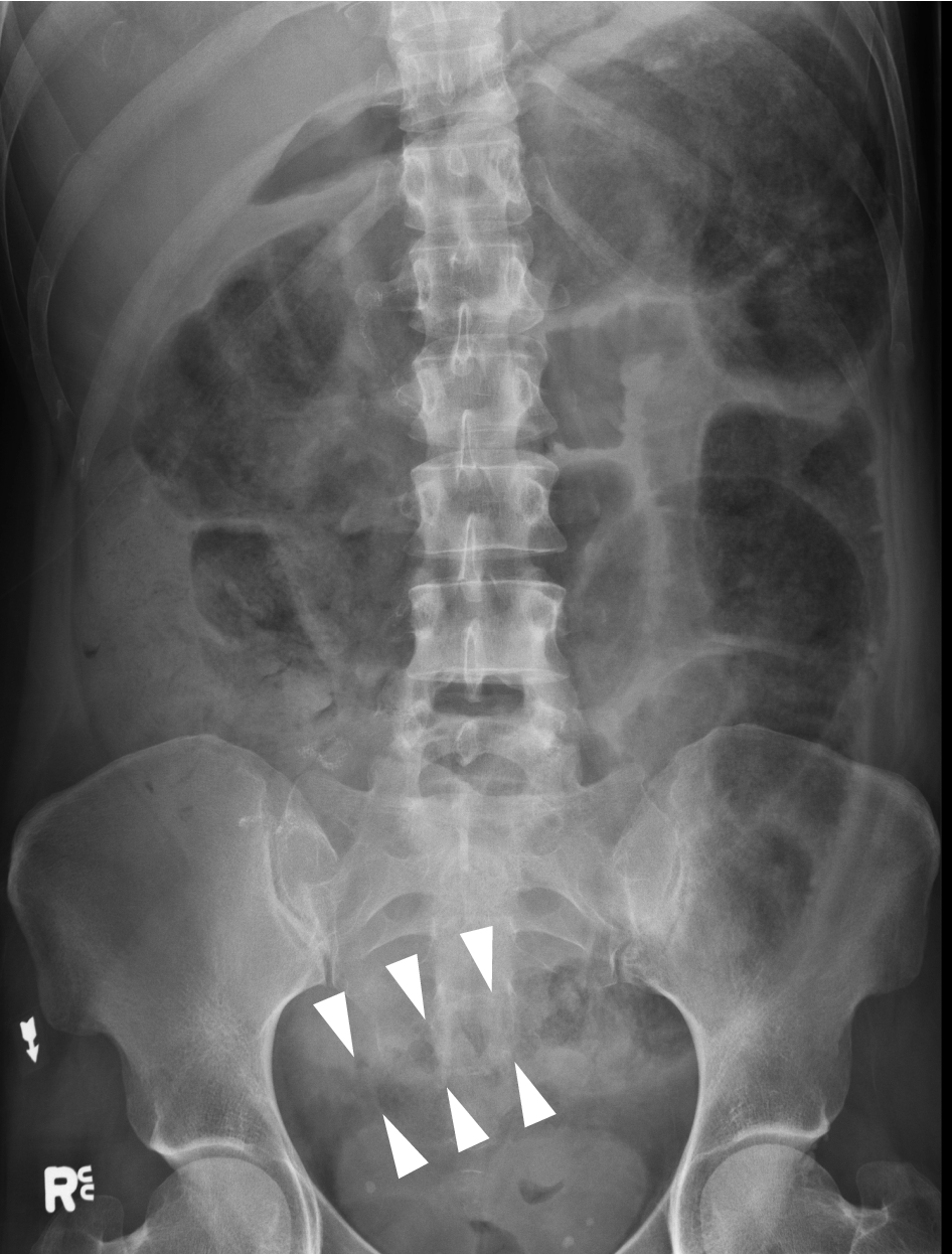 Source: link.springer.com
It is performed with a higher radiation dose and larger dose of IV contrast which helps to evaluate subtle areas of bowel inflammation. Tutorials covering chest X-ray abdominal X-ray and trauma X-ray interpretation. Learn a structured approach to interpreting X-rays. X-rays use beams of energy that pass through body tissues onto a special film and make a picture. This provides an excellent look at the large and small bowel.
 Source: link.springer.com
Abdominal X-Ray - Small bowel obstruction - Small bowel obstruction can be identified by the dilated loops of centrally placed bowel with the venae commitantes circular bands of muscle that span the entire width of the bowel. REM is the term. Because of the difference in X-ray absorption by air and soft tissues the intestinal structures intestinal air can be differentiated from their surroundings. It can also be done to find an object that has been swallowed or to look for a. Send thanks to the doctor.
 Source: link.springer.com
This is a higher quality study than a standard CT. This is a higher quality study than a standard CT. A US doctor answered Learn more. It is performed with a higher radiation dose and larger dose of IV contrast which helps to evaluate subtle areas of bowel inflammation. This is a CT of the Abdomen and Pelvis Enterography protocol.
 Source: youtube.com
They show pictures of your internal tissues bones and organs. Learn a structured approach to interpreting X-rays. Radiology and medical imaging tutorials for medical students and allied health care professionals. This is a higher quality study than a standard CT. Kidney and urinary bladder stones and gallstones.
If you find this site helpful, please support us by sharing this posts to your favorite social media accounts like Facebook, Instagram and so on or you can also save this blog page with the title abdominal x ray tutorial by using Ctrl + D for devices a laptop with a Windows operating system or Command + D for laptops with an Apple operating system. If you use a smartphone, you can also use the drawer menu of the browser you are using. Whether it’s a Windows, Mac, iOS or Android operating system, you will still be able to bookmark this website.


