Prominent renal pyramids ultrasound
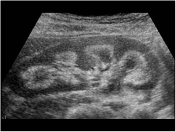
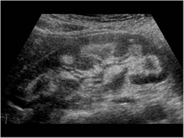
Prominent Renal Pyramids Ultrasound. Should I be concerned about this and what is usually the course of action. Robert Jones RJonesSonoEM Normal medullary. The hypoechoic pyramids normally appear relatively large at this age. They span almost the entire cortex.
 Echogenic Renal Pyramids Differential Radiology Reference Article Radiopaedia Org From radiopaedia.org
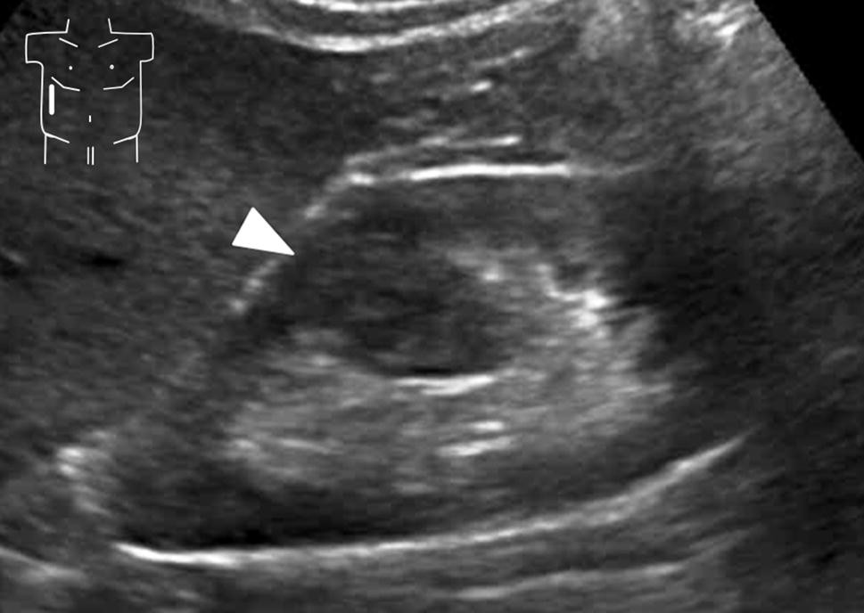
Nephrologists must therefore have a thorough understanding of renal anatomy and the sonographic appearance of normal kidneys and lower urinary tract and they must be able to. They can appear markedly hypoechoic labeled m in image and easily confused as hydro by novice scanners. The renal pelvis may also be seen as a prominent fluid-filled structure when it is extrarenal that is primarily outside of the renal sinus. This may be explained by the larger volume of medulla in the neonatal kidney which results in a ratio of cortex to medulla of 1641 in the neonate as compared with a ratio of 2591 in the adult. Prominent pyramids were present in 50 of subjects with renal cortical echogenicity RCE equal to liver but also in 21 of subjects with RCE less than liver. Prominent pyramids were present in 50 of subjects with renal cortical echogenicity RCE equal to liver but also in 21 of subjects with RCE less than liver.
These normal variations must be differentiated from hydronephrosis.
Place pulse wave Doppler gate interlobar vessels and activate Doppler. The renal pelvis may also be seen as a prominent fluid-filled structure when it is extrarenal that is primarily outside of the renal sinus. They span almost the entire cortex. PAEDIATRIC RENAL ULTRASOUND - Normal. ReadDownload File Report Abuse. Place pulse wave Doppler gate interlobar vessels and activate Doppler.
 Source: pubs.rsna.org
A renal parenchymal thickness under 1 cm is considered abnormal. A renal parenchymal thickness under 1 cm is considered abnormal. Nephrologists must therefore have a thorough understanding of renal anatomy and the sonographic appearance of normal kidneys and lower urinary tract and they must be able to. This may be explained by the larger volume of medulla in the neonatal kidney which results in a ratio of cortex to medulla of 1641 in the neonate as compared with a ratio of 2591 in the adult. They can appear markedly hypoechoic labeled m in image and easily confused as hydro by novice scanners.
 Source: radiopaedia.org
Prominent Renal Pyramids Free PDF eBooks. There is also a noted lack of echogenicity in the renal sinus region in neonates. The kidneys were found to be large in four patients and small in five patients. Posted on January 20 2015. This may relate to recent improvements in ultrasound technology.
 Source: radiopaedia.org
They can appear markedly hypoechoic labeled m in image and easily confused as hydro by novice scanners. Posted on January 20 2015. Takes until 6 months. The hypoechoic pyramids normally appear relatively large at this age. Developmental Renal Pseudotumors Prominent Columns of Bertin Prominent or hypertrophied columns of Bertin also known as septa of Bertin is a normal variant of the kidney formed of hyper-trophied cortical tissue located between the pyramids that projects into the renal sinus re-sulting in splaying of the sinus.
 Source: researchgate.net
Takes until 6 months. Posted on January 20 2015. They can appear markedly hypoechoic labeled m in image and easily confused as hydro by novice scanners. Our study expands the age at which prominently hypoechoic medullary pyramids can be considered a normal finding. Normal sonographic appearance of the renal pyramids in young infants.
 Source: pubs.rsna.org
Medullary sponge kidney is a condition where the collecting tubules in the renal pyramids are ectatic. The hypoechoic pyramids normally appear relatively large at this age. Has prominent medullary pyramids. The Renal pyramids are hyperechoic on ultrasound sometimes with posterior acoustic shadowing. Distal renal tubular acidosis.
 Source: pinterest.com
Renal size was unhelpful in discriminating between different conditions. Normal prominent medullary pyramids on renal POCUS No hydronephrosis. Lower surrounding tissue pressure allows the pelvis to be chronically dilated. There is also a noted lack of echogenicity in the renal sinus region in neonates. Developmental Renal Pseudotumors Prominent Columns of Bertin Prominent or hypertrophied columns of Bertin also known as septa of Bertin is a normal variant of the kidney formed of hyper-trophied cortical tissue located between the pyramids that projects into the renal sinus re-sulting in splaying of the sinus.
 Source: pinterest.de
Nephrocalcinosis of the kidney is the accumulation of calcium in the kidney and in the case that you. Be cautious not to mistake them in the neonate as prominence of the collecting system. The white pyramid sign also known as dense renal medulla sign DRM or hyperdense renal pyramids sign refers to the CT appearance of the medullary pyramids of the kidney which can be seen normally on unenhanced CT scans as high-attenuation triangular structures. A renal parenchymal thickness under 1 cm is considered abnormal. This may be explained by the larger volume of medulla in the neonatal kidney which results in a ratio of cortex to medulla of 1641 in the neonate as compared with a ratio of 2591 in the adult.
 Source: renalfellow.org
They span almost the entire cortex. Prominent pyramids were present in 50 of subjects with renal cortical echogenicity RCE equal to liver but also in 21 of subjects with RCE less than liver. PAEDIATRIC RENAL ULTRASOUND - Normal. Echogenic renal pyramids in children can be due to many different causes. Developmental Renal Pseudotumors Prominent Columns of Bertin Prominent or hypertrophied columns of Bertin also known as septa of Bertin is a normal variant of the kidney formed of hyper-trophied cortical tissue located between the pyramids that projects into the renal sinus re-sulting in splaying of the sinus.
Source: researchgate.net
Developmental Renal Pseudotumors Prominent Columns of Bertin Prominent or hypertrophied columns of Bertin also known as septa of Bertin is a normal variant of the kidney formed of hyper-trophied cortical tissue located between the pyramids that projects into the renal sinus re-sulting in splaying of the sinus. The hypoechoic pyramids normally appear relatively large at this age. Place Color Flow Doppler on the Kidney. Table 3 - Ultrasound. Ultrasound is commonly used in nephrology for diagnostic studies of the kidneys and lower urinary tract and to guide percutaneous procedures such as insertion of hemodialysis catheters and kidney biopsy.
Source:
Our study expands the age at which. The kidneys were found to be large in four patients and small in five patients. Nephrologists must therefore have a thorough understanding of renal anatomy and the sonographic appearance of normal kidneys and lower urinary tract and they must be able to. Has prominent medullary pyramids. The neonatal kidney often demonstrates prominent and hypoechoic renal pyramids.
 Source: ultrasoundcases.info
The hypoechoic pyramids normally appear relatively large at this age. Therefore parenchymal thickness is often easier to measure. Recognize this normal finding. Normal renal pyramids are hypoechoic relative to the renal cortex indepen-Figure 2. Initially in the neonatal period there is almost no medullary fat.
 Source: pubs.rsna.org
Prominent pyramids were present in 50 of subjects with renal cortical echogenicity RCE equal to liver but also in 21 of subjects with RCE less than liver. Table 3 - Ultrasound. Recognize this normal finding. Posted on January 20 2015. Prominent pyramids were present in 50 of subjects with renal cortical echogenicity RCE equal to liver but also in 21 of subjects with RCE less than liver.
 Source: link.springer.com
Normal prominent medullary pyramids on renal POCUS No hydronephrosis. The kidneys were found to be large in four patients and small in five patients. Renal size was unhelpful in discriminating between different conditions. Initially in the neonatal period there is almost no medullary fat. Echogenic renal pyramids in children can be due to many different causes.
 Source: pubs.rsna.org
Anatomically this is secondary to the absence of renal sinus adipose tissue in the kidney of the neonate. Place Color Flow Doppler on the Kidney. Differential diagnosis Nephrocalcinosis Iatrogenic most common cause furosemide frusemide vitamin D. The neonatal kidney often demonstrates prominent and hypoechoic renal pyramids. Takes until 6 months.
 Source: ultrasoundcases.info
Figure 59 demonstrates cortical versus parenchymal thickness measurements. Prominent pyramids were present in 50 of subjects with renal cortical echogenicity RCE equal to liver but also in 21 of subjects with RCE less than liver. Table 3 - Ultrasound. ReadDownload File Report Abuse. UreteralKidney Stones Nephrolithiasis Renal Cysts.
If you find this site value, please support us by sharing this posts to your own social media accounts like Facebook, Instagram and so on or you can also bookmark this blog page with the title prominent renal pyramids ultrasound by using Ctrl + D for devices a laptop with a Windows operating system or Command + D for laptops with an Apple operating system. If you use a smartphone, you can also use the drawer menu of the browser you are using. Whether it’s a Windows, Mac, iOS or Android operating system, you will still be able to bookmark this website.


