Sheep brain ventral view
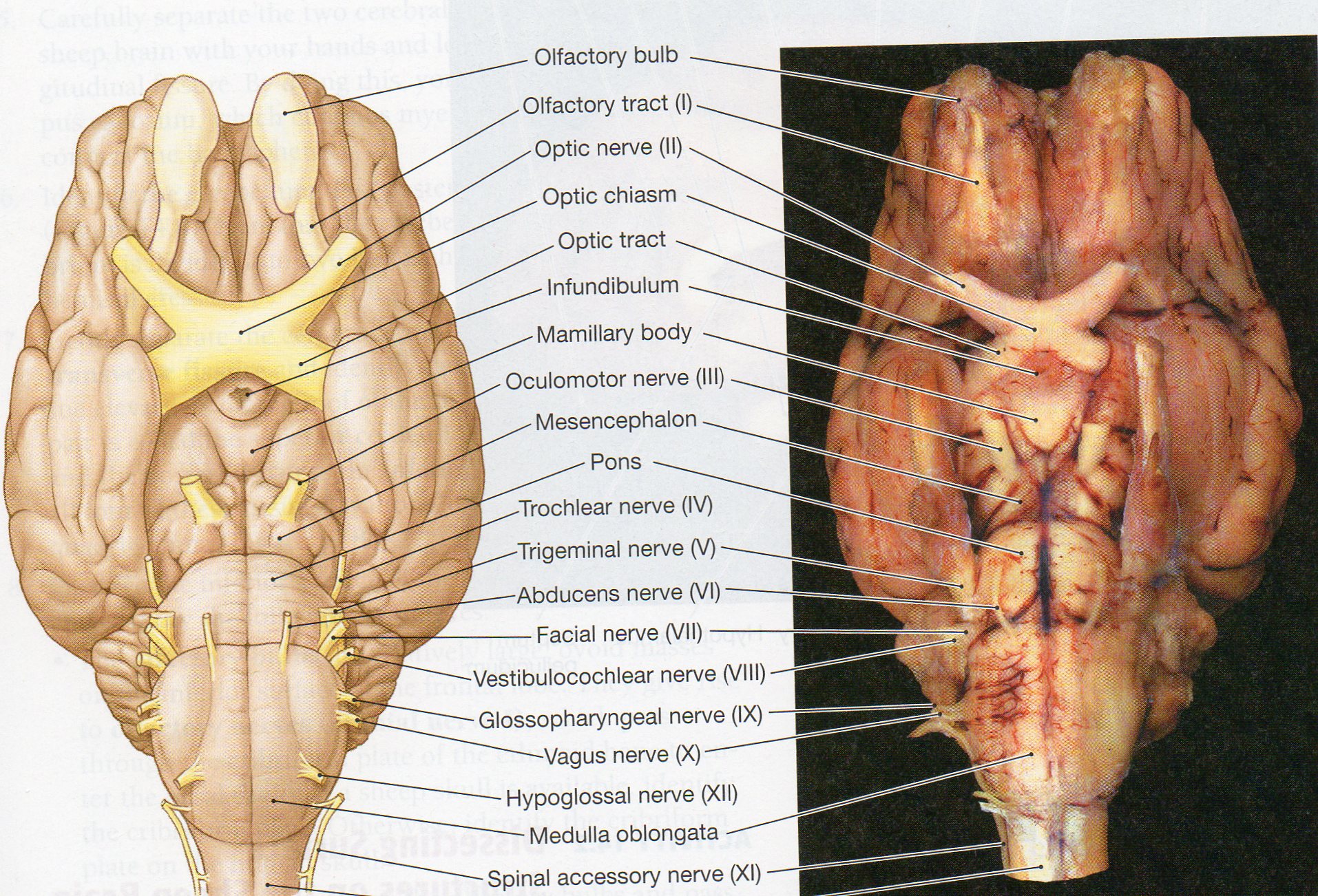
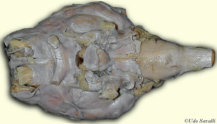
Sheep Brain Ventral View. Various surface components of the rhinencephalon are labeled on the left side. First image in each group of 3. Sheep Brain Dissection Ventral View Orange pin. Ventral view with dura.
 Sheep Brain From bp-web.baypath.edu
Sheep Brain Dissection Ventral View Orange pin. Olfactory Blub only one other cut off-The Olfactory bulb contains nerves that deal with olfaction smell. To View The Nerves Highlighted by Color. Between these the orange pic is in the lumen of the pituitary stalk infundibulum. Ventral View of the Sheep Brain. Pons -Pons serve as.
Sheep Brain Dissection Guide 3.
To View The Nerves Highlighted by Color. Various surface components of the rhinencephalon are labeled on the left side. The most prominent structure visible on the ventral side of the sheep brain is half of the optic chiasma which is where the two optic nerves cross over each other and form an X shape. Ventral View of the Sheep Brain. 6 Dorsal View of the Sheep Brain 7. You may have removed the optic removed the chiasma with the dura mater.
 Source: pinterest.com
Between these the orange pic is in the lumen of the pituitary stalk infundibulum. You can still see some structures on the brain before you remove the dura mater. Removal of the Dura Mater. Double click on a nerve to produce a table with important information about that nerve. Find the optic chiasma half on your brain.
 Source: courses.lumenlearning.com
The optic chiasm green pic marks the rostral end of the hypothalamus optic nerves are rostral and optic tracts are caudal to the chiasmMamillary bodies red mark the caudal end of the hypothalamus. Sheep brain ventral view 1. Longitudinal Fissure-The longitudinal fissure divides the brain into two halves called the left and right brain. Sheep Brain Atlas An ongoing project UCLA Department of Psychology Psychology 116 Website by Kevin Noguchi. Removal of the Dura Mater.
 Source: slidetodoc.com
Midbrain connects midbrain with cerebrum. Anatomy of the sheep brain video for anatomy class - practice for the practical exam. Various surface components of the rhinencephalon are labeled on the left side. 6 Dorsal View of the Sheep Brain 7. Sheep brain ventral view 1.
Source: imagequiz.co.uk
Several important parts of the visual system are visible in the ventral view of the brain. Examine the ventral surface of the sheep brain. There is a printable worksheet available for download here so you can take the quiz with pen and paper. These two structures will likely be pulled off when you remove the dura mater. This image shows the ventral.
 Source: courses.lumenlearning.com
The sheeps brain has a more developed olfactory bulb when compared to the human brain. Ventral View Sheep Brain. External Views. The human brain has a larger frontal lobe than the sheeps brain. Identify the olfactory bulb 2 piriform lobe 7 and lateral olfactory tract 6.
Source: csun.edu
There is a printable worksheet available for download here so you can take the quiz with pen and paper. Ventral View of the Sheep Brain. Learn vocabulary terms and more with flashcards games and other study tools. Pons -Pons serve as. Ventral view with dura.
 Source: www2.palomar.edu
Anatomy of the sheep brain video for anatomy class - practice for the practical exam. The optic chiasm green pic marks the rostral end of the hypothalamus optic nerves are rostral and optic tracts are caudal to the chiasmMamillary bodies red mark the caudal end of the hypothalamus. Longitudinal Fissure-The longitudinal fissure divides the brain into two halves called the left and right brain. 15M ratings 277k ratings See thats what the app is perfect for. Midbrain connects midbrain with cerebrum.
 Source: quizlet.com
The human brain has a larger frontal lobe than the sheeps brain. To View The Nerves Highlighted by Color. You may have removed the optic removed the chiasma with the dura mater. There is a printable worksheet available for download here so you can take the quiz with pen and paper. Sounds perfect Wahhhh I dont wanna.
 Source: bp-web.baypath.edu
Cerebral hemispheres frontal cortex Mammillary bodies. Pons -Pons serve as. Examine the ventral surface of the sheep brain. There is a printable worksheet available for download here so you can take the quiz with pen and paper. Oculomotor Nerve which has 3 main motor functions which are.
 Source: personal.psu.edu
Double click on a nerve to produce a table with important information about that nerve. Pons -Pons serve as. Brain with Dura Mater Intact. Between these the orange pic is in the lumen of the pituitary stalk infundibulum. Also the olfactory peduncle 3 and the medial olfactory tract 4 are labeled.
 Source: quizlet.com
Sheep Brain Dissection Elza Joseph and Nias sheep brain dissection portfolio. Brain with Dura Mater Intact. Take special note of the pituitary gland and the optic chiasma. The sheep brain is enclosed in a tough outer covering called the dura mater. In this image a student is bending the cerebellum down to show the superior and inferior colliculi.
 Source: savalli.us
Sheep Brain Atlas An ongoing project UCLA Department of Psychology Psychology 116 Website by Kevin Noguchi. Labels added to arrows. External Views. Several important parts of the visual system are visible in the ventral view of the brain. Spinal cord Anterior Close-Up View Posterior Close-Up View.
 Source: amherst.edu
The next several steps will view this surface of the brain. Cerebral hemispheres frontal cortex Mammillary bodies. The human brain and sheep brain have the major difference that humans can think write invent or create with their brains whereas sheep. In this image a student is bending the cerebellum down to show the superior and inferior colliculi. The human brain is rounded whereas the sheeps brain is elongated in shape.
 Source: pinterest.com
Hypothalamus olfactory relay station. The sheeps brain has a more developed olfactory bulb when compared to the human brain. The left brain is associated with logical thinking while the right brain is associated. Take special note of the pituitary gland and the optic chiasma. Back Home Ventral Structures Labelled.
Source: shefalitayal.com
External Views. 6 Dorsal View of the Sheep Brain 7. Ventral View of the Sheep Brain. Spinal cord Anterior Close-Up View Posterior Close-Up View. External Views.
If you find this site beneficial, please support us by sharing this posts to your own social media accounts like Facebook, Instagram and so on or you can also save this blog page with the title sheep brain ventral view by using Ctrl + D for devices a laptop with a Windows operating system or Command + D for laptops with an Apple operating system. If you use a smartphone, you can also use the drawer menu of the browser you are using. Whether it’s a Windows, Mac, iOS or Android operating system, you will still be able to bookmark this website.


